
VRT (Volume Rendering Technique) is used to display a 2D projection of a 3D discretely sampled data set. It provides a clearer depiction of the complex anatomy which can be used for surgical planning or during discussions with referring physicians.
Accurate pulmonary nodule detection is a crucial step in lung cancer screening. Computer-aided detection (CAD) systems are not routinely used by radiologists for pulmonary nodule detection in clinical practice despite their potential benefits. Maximum intensity projection (MIP) images improve the detection of pulmonary nodules in radiological evaluation with computed tomography (CT) scans.
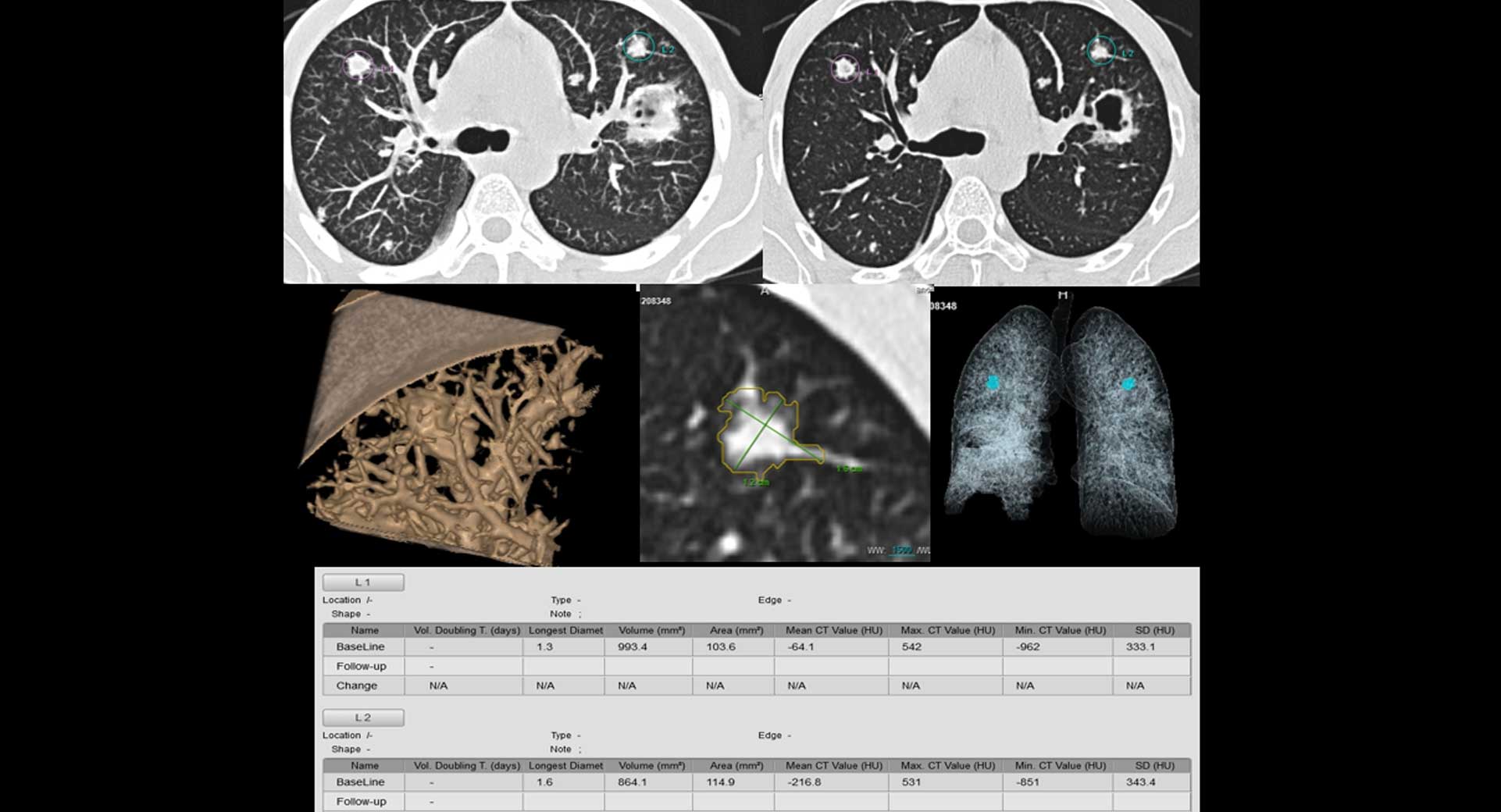
Nodule detection, segmentation and measurement assistance. Nodule quantitative analysis. Follow-up analysis. One-step structured report
Lung density, measured as percentage of lung volume with attenuation −750 to −900 HU (%HDS) for never-smokers (NS), smokers (S), COPD current smokers
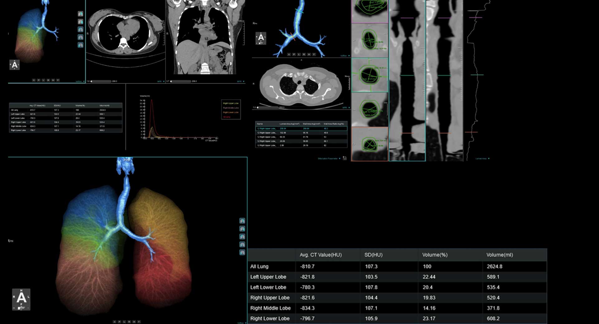
Lung Extraction and Contour Editing.
•Pulmonary Density and Volume Measurement.
•Emphysema Ratio Quantification.
•Quantitative Data Analysis and Export
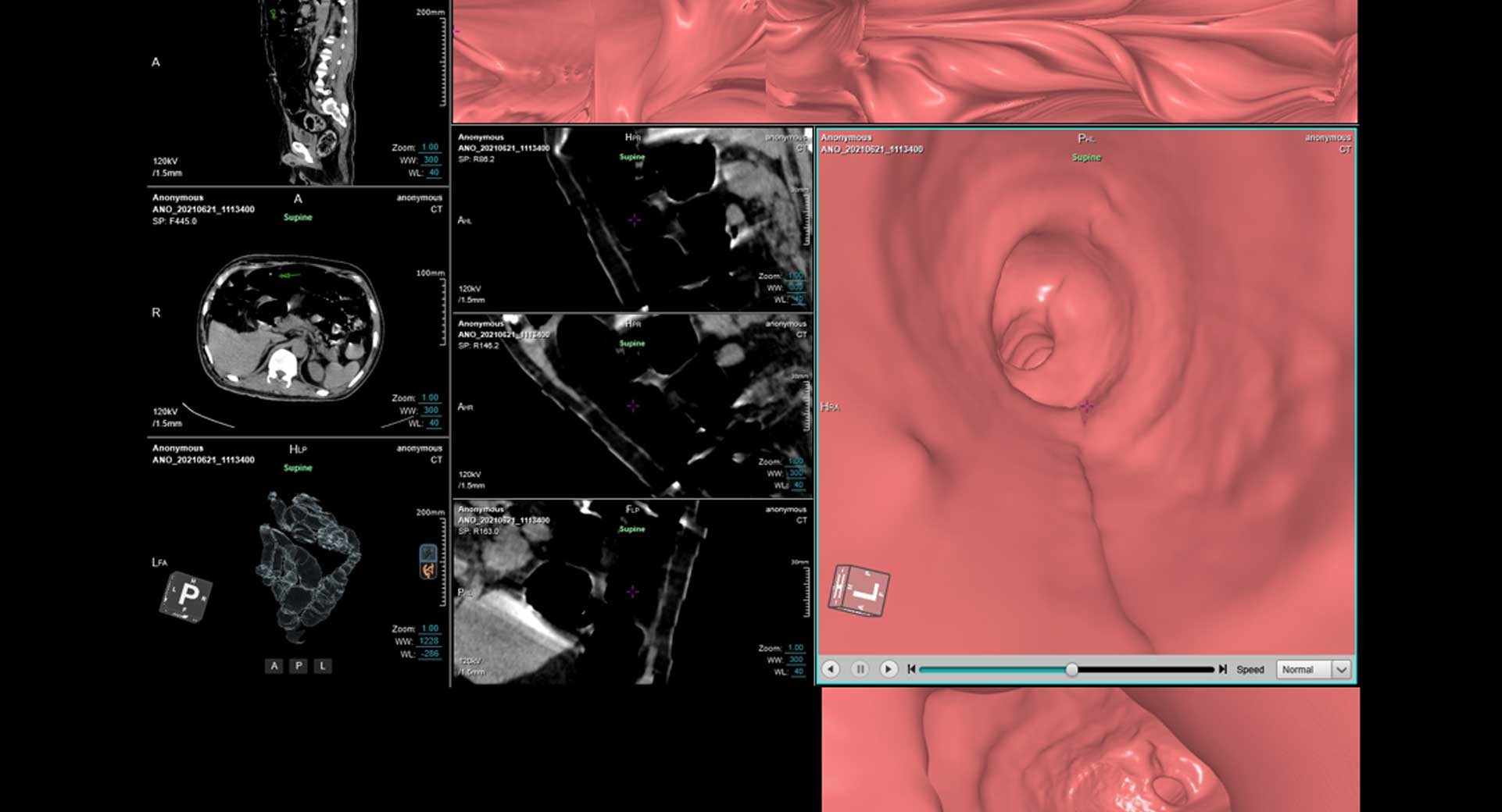
A colonoscopy (koe-lun-OS-kuh-pee) is an exam used to detect changes or abnormalities in the large intestine (colon) and rectum. During a colonoscopy, a long, flexible tube (colonoscope) is inserted into the rectum. A tiny video camera at the tip of the tube allows the doctor to view the inside of the entire colon
•Automatic colon segmentation.
•Electronic colon cleansing.
•Virtual endoscopy.
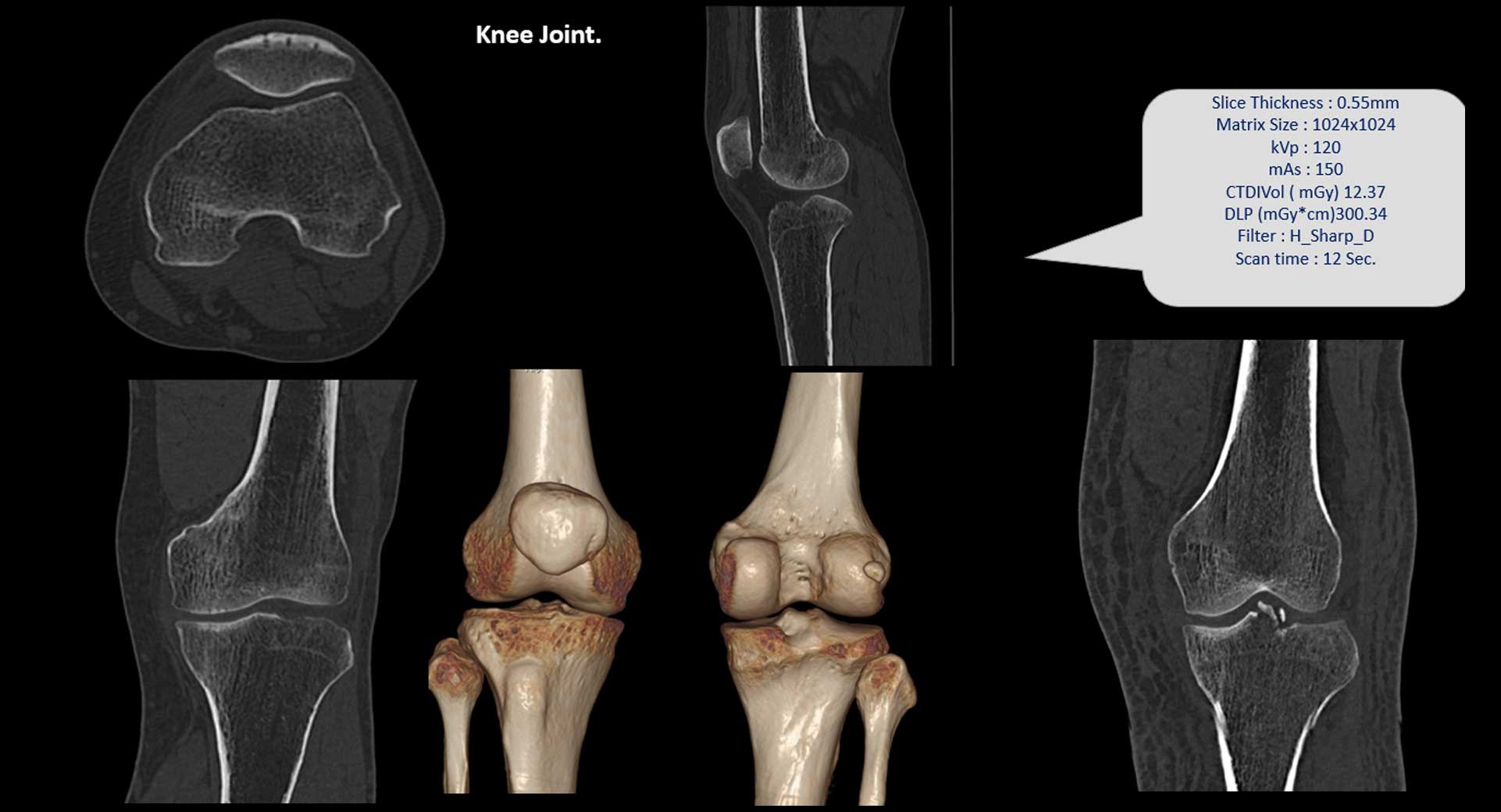
A knee X-ray can help find the cause of pain, tenderness, swelling, or deformity of the knee. It can show broken bones or a dislocated joint
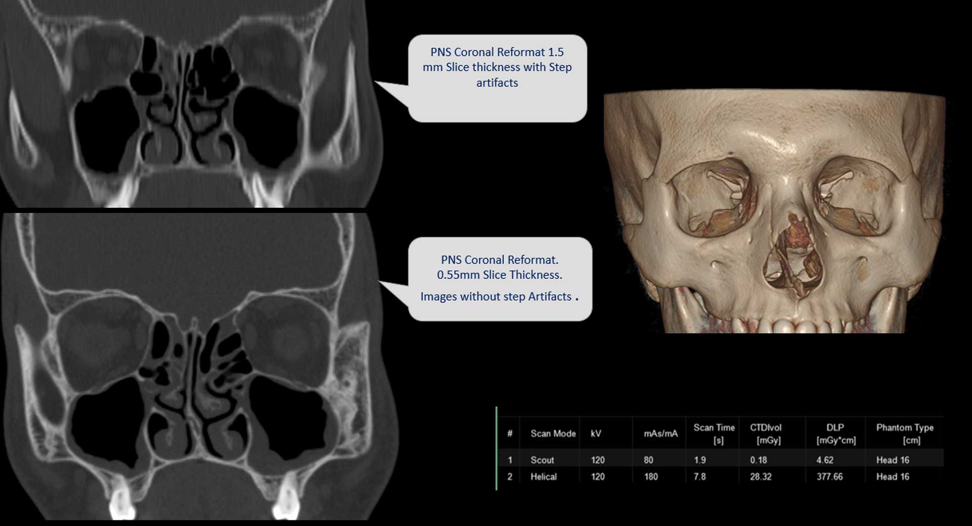
PNS (Para-Nasal Sinus) is an imagining test that is used to visualize details of the PNS. The test may refer to sinus X-Ray or X-Ray of paranasal sinus radiography. The X-Ray PNS test is usually performed with little to minimal pain or discomfort. X-Ray uses a small amount of radiation and is not to be performed during pregnancy.
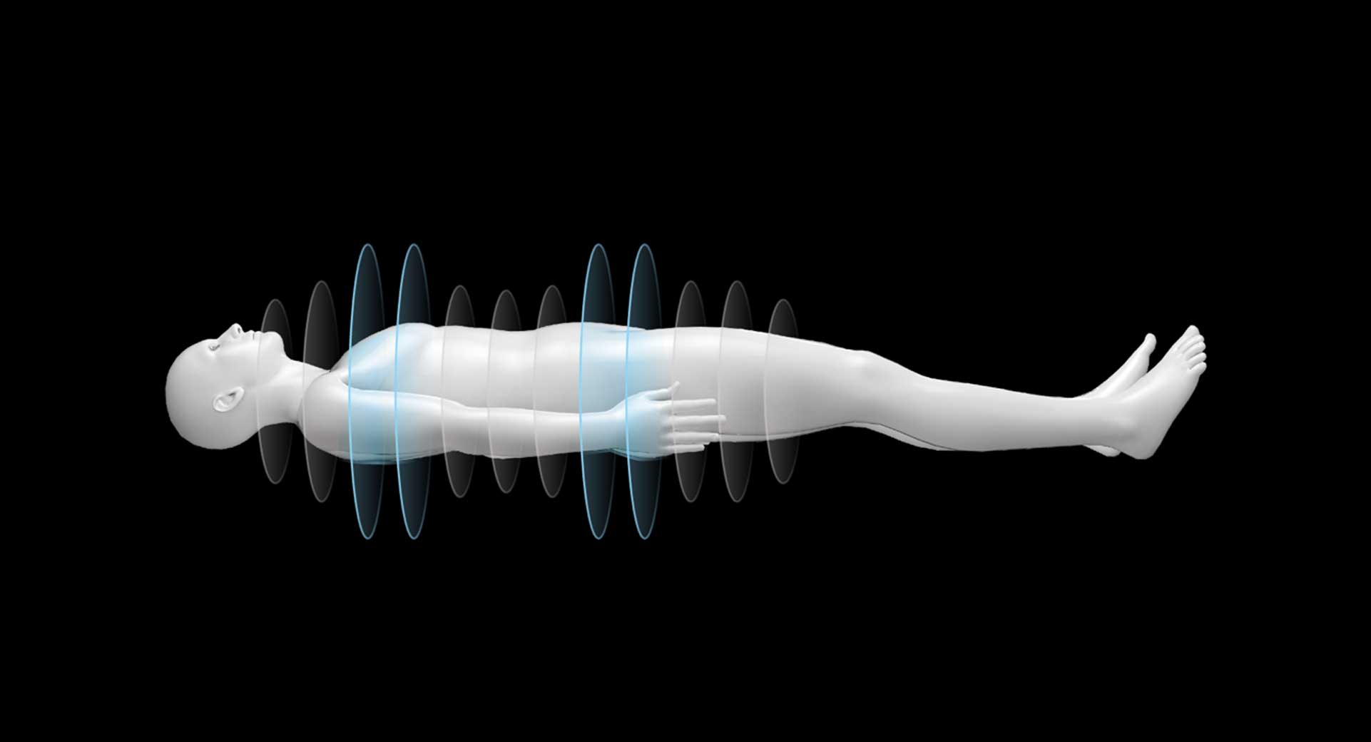
With helical CT, the patient is moved through a rotating x-ray beam and detector set. From the perspective of the patient, the x-ray beam from the CT traces a helical path. The helical path results in a three-dimensional data set, which can then be reconstructed into sequential images for an image stack.
Helical CT allows a scan to be performed in a single breath-hold.
Most modern CT protocols use helical acquisition due to its speed and because it reduces misregistration from patient movement or breathing. Sequential scanning (step-and-shoot) acquisition is still used in some situations (e.g. helical acquisition can lead to artifacts on head CT).
Based on anatomic structure information, it generates optimized 3D dose distribution plan.
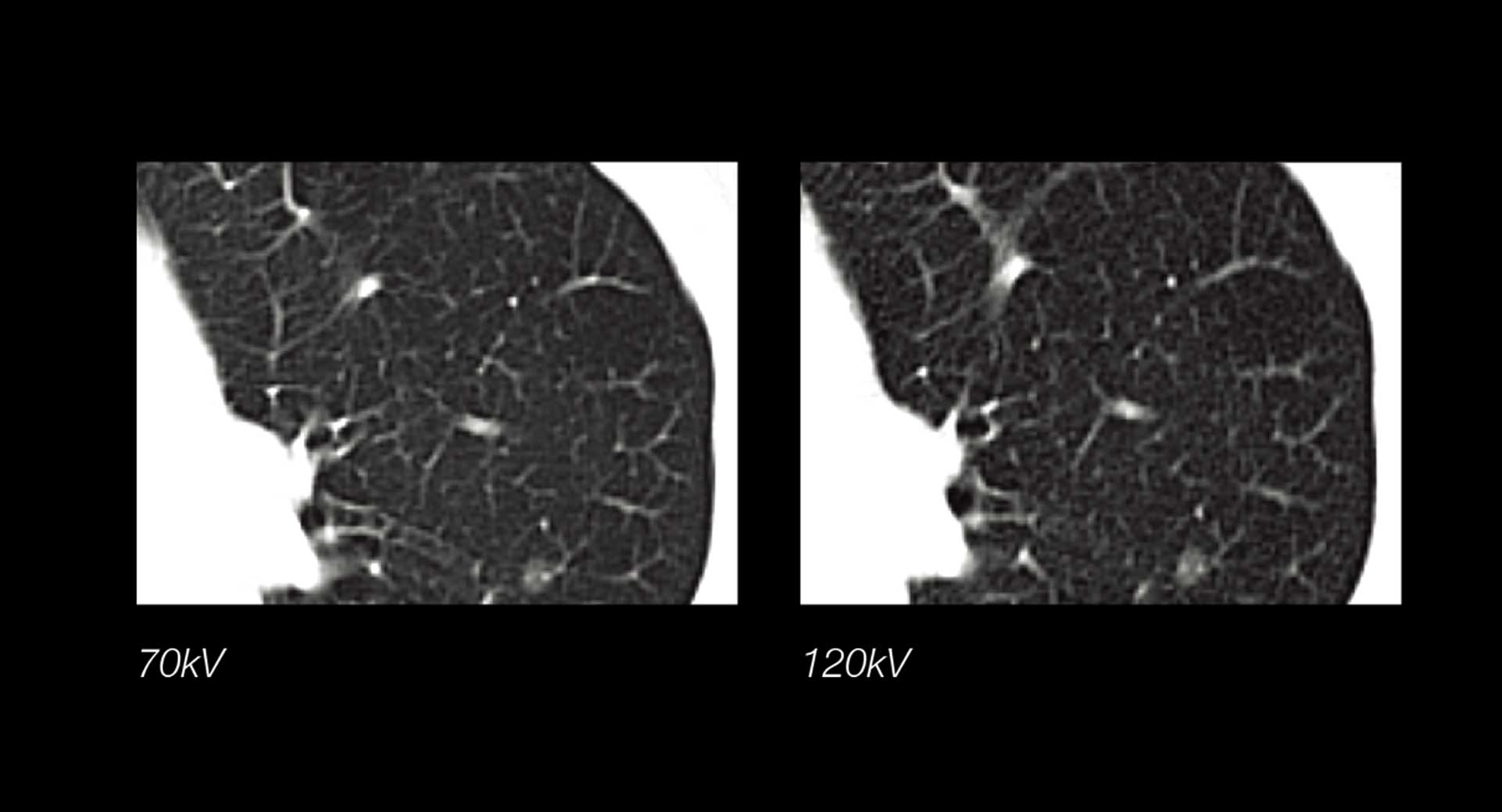
70kV Dose reduction can increase SNR for Iodine-based contrast agents especially for small BMI or younger patients.
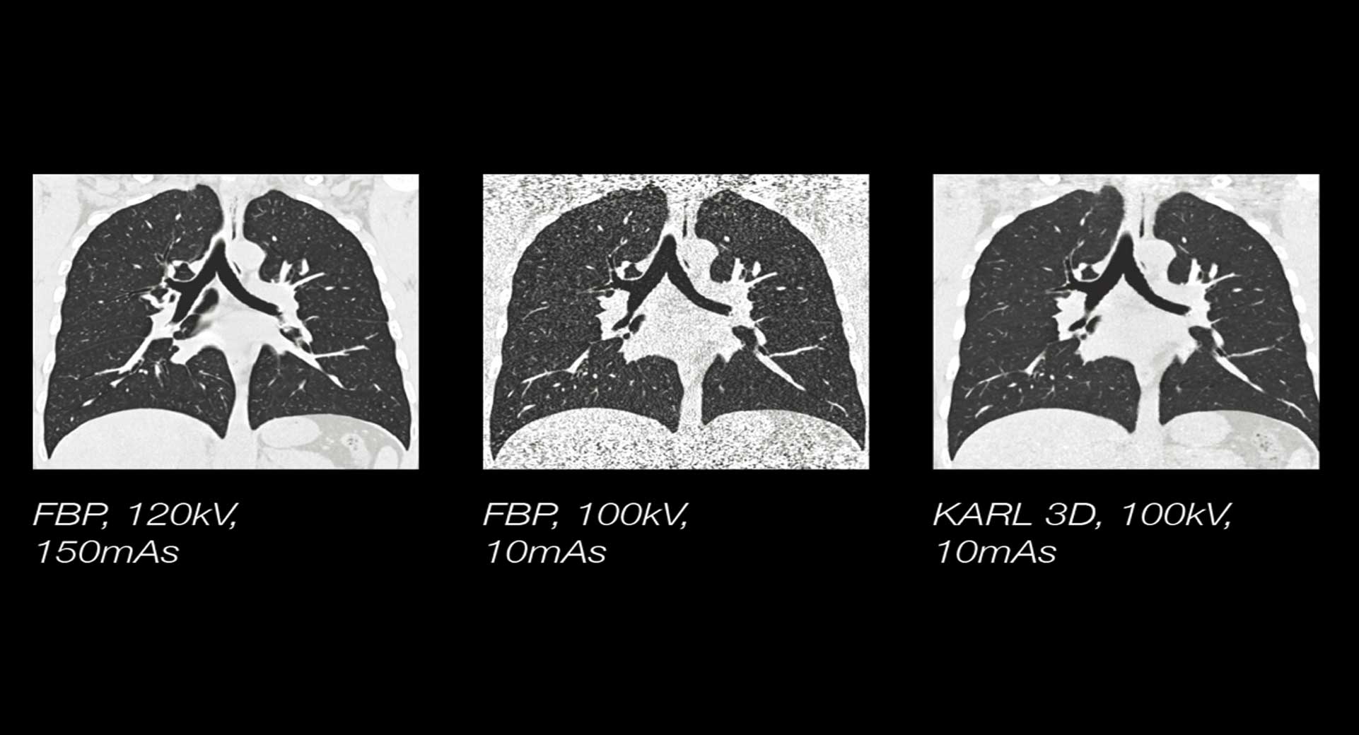
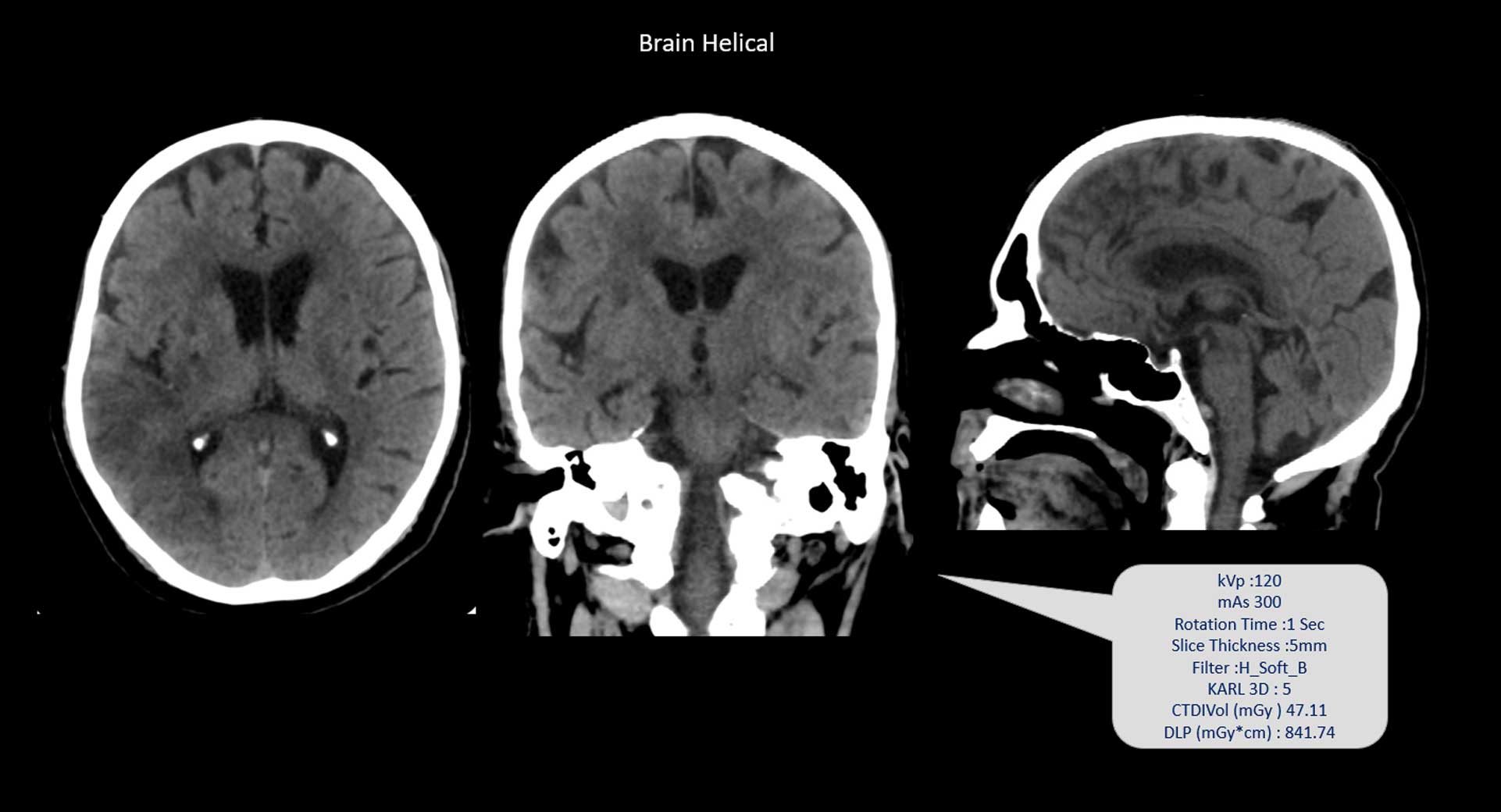
KARL 3D employs a generalized gradient descent iterative denoising technique in both projection and image spaces. The unique KARL 3D Iterative Reconstruction Technology allows users to reduce the radiation dose significantly without sacrificing image quality.
The intuitive and intelligent workflow boosts efficiency and effectiveness of routine practices. Along with the unique Z-Detector, reliable components and multiple integrated advanced applications, we brings youmore access to advanced technologies.Intelligently plans the exam for individual patients. Clinical parameters are automatically adapted to the needs of the particular patient.The system anticipates the operator’s needs and prepares itself to reduce delays. Minimizes the wait time between operation procedures and accelerates CT exam workflow significantly.